
|

|


|
|
|
|
|
|
|
|
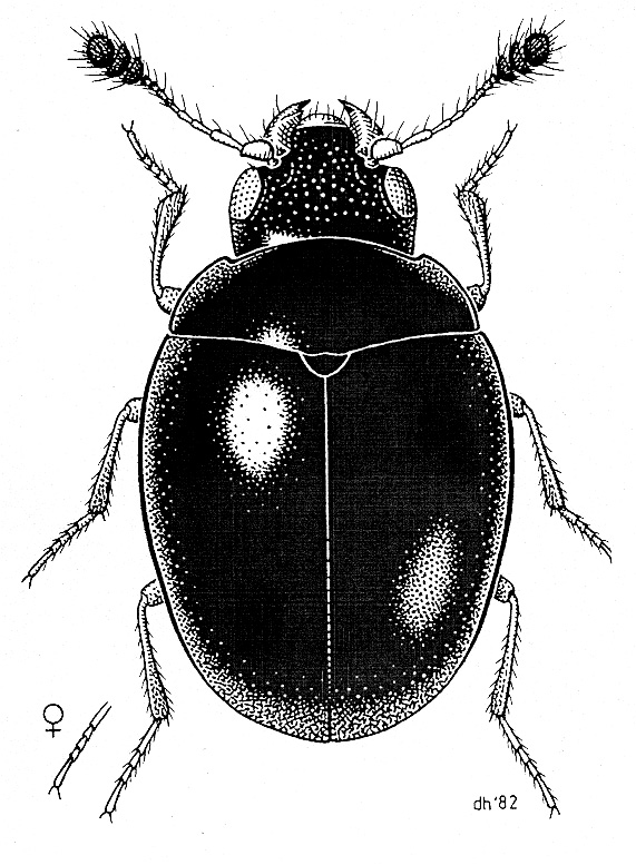 Cyclaxyra politula
Drawing by D.W.Helmore |
M.L. Gimmel, R.A.B. Leschen, S.A. Slipinski. 2009.
Review of the New Zealand endemic family Cyclaxyridae, new family (Coleoptera: Polyphaga).
Acta Entomologica Musei Nationalis Pragae, 2009, 49 (2): 511-528.
По данным статьи единственный род семейства содержит всего 2 эндемичных для Новой Зеландии вида - C. politula (Broun, 1881) (= C. impressa Broun, 1915) и C. jelineki sp. n. Пока полный текст статьи недоступен - мы приводим ниже описание семейства из руководства на компакт-диске (Lawrence et al., 1999).
А.Л. Лобанов, сентябрь 2010 г.
В 2013 году в нашей библиотеке появилась полная копия статьи (Gimmel et al., 2009).
Литература
Broun, T. 1881. Manual of the New Zealand Coleoptera. Part II. Colonial Museum and Geological Survey Department, Wellington, pp. xxi-xxiii + 653-744.
Broun, T. 1893. Manual of the New Zealand Coleoptera. Parts V., VI., VII. New Zealand Institute, Wellington, pp. v-xvii + 975-1504.
Crowson, R. A. 1952. The classification of the families of British Coleoptera (part). Entomologist's Monthly Magazine 88: 109-132 (Cucujoidea: Clavicornia).
Klimaszewski, J. and J. C. Watt, 1997. Fauna of New Zealand. Number 37. Coleoptera: Family Group Review and Keys to Identification. Manaaki Whenua Press, Lincoln, Canterbury, 199 pp. Wellington. 56 pp.
Lawrence J.F., A.M. Hastings, M.J. Dallwitz, T.A. Paine & E.J. Zurcher, 1999. Beetles of the World: A Key and Information System for Families and Subfamilies. CD-ROM, Version 1.0 for MS-Windows. Melbourne: CSIRO Publishing.
Sen Gupta, T. and R. A. Crowson, 1966. A new family of cucujoid beetles, based on six Australian and one New Zealand genera. Annals and Magazine of Natural History, (13) 9: 61-85.
Watt, J. C. and R. A. Crowson, 1986. Cyclaxyra Broun, 1893 (Insecta, Coleoptera): proposed conservation by the suppression of Melanocrhoa Broun, 1882. Z.N.(S.)2511. Bulletin of Zoological Nomenclature 43: 196-198.
Wu H., Li L., Ding M. 2018.
The first cyclaxyrid beetle from Upper Cretaceous Burmese amber (Coleoptera: Cucujoidea: Cyclaxyridae).
PDF-file in our library
CYCLAXYRIDAE
Classification. Polyphaga: Cucujoidea.
Distribution. The genus Cyclaxyra occurs only in New Zealand.
Biology. Cyclaxyra adults and larvae have been collected on bark in masses of sooty mold (Ascomycetes: Capnodiaceae etc.).
General appearance. Total length 1.5-2 mm. Ratio of body length to greatest body width 1.25-1.35. Body strongly convex. Sides of body evenly curved. Body not capable of conglobation (rolling into a ball). Upper surfaces of body glabrous or subglabrous. Vestiture of upper surfaces not including stiff, erect, dark bristles. Vestiture of upper surfaces not including scales or scale-like setae. Upper surfaces of body without deep foveae. Prothorax, metathorax and-or abdomen without extrusible glands. Underside of body without hydrofuge surface(s).
Head. Head width just behind eyes not distinctly greater than prothoracic width. Head not or slightly declined. Head not entirely concealed from above by pronotum. Head without elongate rostrum. Head not abruptly constricted posteriorly. Temples absent or not closely adpressed to prothorax. Temples absent. Transverse occipital ridge or carina absent. Occiput without stridulatory file. Head without ocelli. Compound eyes present. Eyes not or only slightly protuberant. Eyes finely facetted. Eyes without interfacetal setae. Ommatidium of the acone type. Eye entire. Anterior or mesal edge of eye not or only barely emarginate. Posterior edge of eye not or barely emarginate. Antennal insertions exposed from above. Antennal insertions moderately to widely separated. Antennae not borne on raised tubercles. Subantennal groove or cavity on head well developed. Frontoclypeal suture absent or incomplete. Clypeus not laterally emarginate. Anterior edge of clypeus or clypeolabrum straight to convex. Mouth cavity anteriorly or anteroventrally oriented. Pregular area without laterally opening cavities. Head ventrally without paired subgenal ridges. Head without anteriorly-projecting genal processes. Gular sutures widely separated or absent. Corporotentorium narrow. Corporotentorium without median process. Cervical sclerites present. Antennae. Number of antennomeres 11. Antennae when posteriorly extended reaching beyond middle of prothorax but not middle of elytra. Antennae capitate. Antennomeres 3, 4 or 5 to 10 without or with single rami (uniramose). Antennae at least partly pubescent or with obvious modifications. Antennal modifications beginning on antennomere 5 or beyond. First antennomere (scape) less than 3 times as long as 2nd (pedicel). Antenna not geniculate. Antenna with a distinct club. Antenna with strong apical club. Antennal club 3-segmented. Antennal club not 5-segmented or with 2nd segment subequal to or larger than 1st. Antennal club loose. Antennal club not lamellate. Antennal club not or slightly serrate. Antennal club not or slightly flattened. Antennal club not preceded by a cupule. Mouthparts. Labrum at least partly visible. Labrum free, membranous or separated by suture. Major portion of labrum strongly transverse. Apex of labrum subtruncate to slightly convex; or strongly convex, narrowly rounded or acute. Labrum moderately to heavily sclerotized, except at base and-or apex. Mouthparts not forming a piercing or sucking tube. Mandibles present. Mandible short and broad. Mandibular apex moderately to strongly, gradually curved mesally. Mandibular apex bidentate or bilobed. Dorsal part of mandible without tubercle. Dorsal part of mandible without setose cavity. Incisor edge of mandible simple. Mandible with well developed mola. Mandible with well developed prostheca. Prostheca absent or without articulated, sclerotized process. Maxilla with distinct galea and lacinia. Maxillary lobe(s) not stylet-like. Apex of galea or maxillary lobe densely setose or spinose. Apex of galea or maxillary lobe without heavily sclerotized teeth or hooks. Lacinia without hook(s) or spine(s). Apical maxillary palpomere cylindrical to fusiform. Apical maxillary palpomere at least as wide as or longer than preapical one. Maxillary palp without complex palp organ. Apical labial palpomere cylindrical to fusiform. Ligula undivided or finely cleft.
Prothorax. Ratio of pronotal length to greatest pronotal width 0.35-0.4. Prothorax widest posteriorly. Sides of prothorax more or less straight; or moderately to strongly curved. Prothorax not laterally compressed to form cavities for legs. Sides of prothorax moderately to strongly, obliquely or vertically explanate. Base of prothorax not or slightly narrower than elytral bases. Greatest prothoracic width distinctly narrower than greatest elytral width. Lateral pronotal carinae complete. Lateral pronotal carinae simple. Lateral pronotal carinae visible for their entire lengths from above. Lateral pronotal carinae with a raised margin or narrow bead. Lateral portion of prothorax without deep pit. Pronotum without anterolateral callosities. Anterior angles of pronotum distinctly produced forward. Anterior angles of pronotum produced and narrowly rounded or acute. Posterior angles of pronotum obtuse or right. Posterior angles of pronotum not produced and acute. Posterior edge of pronotum more or less straight or evenly rounded. Posterior edge of pronotum simple. Posterior edge of pronotum not or vaguely margined. Discal carinae of pronotum absent. Pronotal disc without paired basal impressions. Pronotum without median longitudinal groove or line. Hypomeron without pit. Anterior portion of prosternum at midline shorter than prosternal process. Lateral portion of prosternum in front of coxae shorter than mid length of procoxal cavity. Anterior edge of prosternum distinctly produced forming chin piece. Prosternum in front of coxae flat to moderately convex. Prosternum in front of coxae without paired lines or carinae. Anterior edge of prosternum without mesal excavation. Anterolateral or ventrolateral portions of prothorax without cavities or grooves. Prothoracic cavities absent. Prothoracic grooves absent. Prosternal process complete. Prosternal process gradually expanded or narrowed and then expanded. Prosternal process flat, concave, or only slightly elevated or curved behind coxae. Prosternal process slightly overlapping mesoventrite; or moderately to strongly overlapping mesoventrite. Apex of prosternal process broadly rounded, angulate or truncate. Prosternal process without transverse groove. Accessory (mesal) procoxal articulation absent. Ventral portion of prothorax on each side with notosternal suture only. Propleuron not extending to anterior edge of prothorax. Propleuron or pleurotrochantin not extending behind coxa. Procoxae not or slightly projecting below prosternum. Procoxa without or with short concealed lateral extension. Procoxal cavities present, procoxae countersunk. Procoxal cavity strongly transverse. Procoxal cavities at middle moderately to widely separated. Procoxal cavities externally open. Procoxal cavities externally broadly open. Postcoxal projection absent or very short. Procoxal cavities without narrow lateral extensions. Procoxal cavities internally open. Prothoracic trochantin or pleurotrochantin at least partly exposed. Promesothoracic clicking mechanism absent.
Elytra. Elytra present. Ratio of elytral length to greatest elytral width 1-1.05. Ratio of elytral length to pronotal length 3.3-3.6. Elytra apunctate, irregularly punctate, or with 5 or fewer distinct puncture rows or striae. Elytra without scutellary striole. Sutural stria absent or not deeply impressed near apex. Abdominal tergites exposed by elytra none or apex of 1. Elytral apices meeting or almost meeting at the suture. Elytral suture not deflected near apex. Elytral apex without internal interlocking tongue. Epipleuron absent or incomplete. Epipleuron not or gradually narrowed. Lateral edge of elytron straight or weakly sinuate.
Pterothorax. Scutellum well developed. Scutellum abruptly elevated. Scutellum anteriorly simple. Scutellum posteriorly broadly rounded or obtusely angulate. Mesoscutum without stridulatory file. Mesoventrite with paired procoxal rests. Paired mesoventral procoxal rests vertical. Anterior edge of mesoventrite without prosternal rest. Mesoventrite not divided by longitudinal groove or discrimen. Anterior edge of mesoventrite at midline on same plane as metaventrite. Mesoventral cavity absent. Mesocoxa not conical and projecting. Mesocoxal cavities present, mesocoxae countersunk. Mesocoxal cavities at middle moderately to widely separated. Mesocoxae separated by more than shortest diameter of coxal cavity. Mesocoxal cavities circular to slightly transverse. Mesocoxal cavities not or slightly oblique. Mesoventrite separated by complete sutures from mesepisterna. Mesepisterna distinctly separated at midline. Mesepisterna without deep pockets. Mesepimeron not visible from above. Mesocoxal cavities open laterally. Mesocoxal cavities not partly closed by metepisterna. Mesoventral process absent or not extending to middle of mesocoxal cavity; or extending at least to middle of mesocoxal cavity. Mesometaventral junction an anteriorly curved, angulate or acute line. Metaventral discrimen or median line absent. Paired postcoxal lines of metaventrite absent. Postcoxal lines of metaventrite absent. Metaventrite longer than first abdominal ventrite. Postcoxal pits of metaventrite absent. Metaventrite flat to slightly convex. Transverse groove of metaventrite absent. Anterior edge of metaventrite without transverse carina between mesocoxal cavities. Exposed portion of metepisternum very long and narrow or absent. Metacoxae contiguous or narrowly separated. Metacoxae not extending laterally to meet elytra or sides of body. Metacoxae completely separated from metaventrite by suture. Metacoxal plates absent. Metacoxae not greatly enlarged. Metacoxae horizontally oriented. Lateral arms of metendosternite moderately to very long. Metendosternal laminae reduced. Ventrolateral processes of metendosternite absent or weakly developed. Anterior process of metendosternite short or absent. Anterior tendons of metendosternite widely separated but not on lateral arms. Apical portion of metendosternite not or only slightly emarginate.
Hind wing. Hind wing well developed. Hind wing with normal transverse folds. Radial cell of hind wing highly reduced or absent. Radial cell of hind wing incomplete or absent. Ratio of length of apical area to total wing length greater than 0.5. Medial bar of hind wing not crossed by fold. Free veins in medial area of hind wing 3 or fewer. Oblongum cell of hind wing absent. Medial fleck of hind wing absent. Medial fleck of hind wing absent or not partly bisected by a vein. Wedge cell of hind wing absent. Anal lobe of hind wing present. Posterior edge of hind wing without fringe of long hairs.
Legs. Femoral attachment of mid trochanter strongly oblique with base of femur separate from coxa. Mesotrochanter not reduced or concealed from below. Metafemur not much wider than mesofemur. Mesotibia not strongly widened. Outer edge of mesotibia simple, crenulate or denticulate. Outer subapical edge of mesotibia without antenna cleaner. Preapical surfaces of mesotibia without ridges or combs. Outer apical angle of mesotibia simple or slightly produced, without lobe, teeth or spines. Mesotibial spurs glabrous or absent. Mesotarsus with 5 distinct tarsomeres (pentamerous). Tarsomeres on hind leg at least as many as on mid leg; or 1 fewer than on mid leg. Tarsomeres on fore leg at least as many as on mid leg. Mesotarsomere 1 well developed and visible. Preapical mesotarsomeres together longer than apical one. Penultimate mesotarsomere not distinctly shorter than antepenultimate. Ventral mesotarsal lobes absent. Mesotarsal claws paired. Mesotarsal claws subequal in length and similar in form and angle of inclination. Mesotarsal claws simple. Appendage on each tarsal claw absent. Mesotarsal claws without setae near base. Mesotarsal empodium absent or with 2 or fewer setae. Inner subapical edge of protibia without antenna cleaner. Hind legs without swimming hairs. Preapical surfaces of metatibia without ridges or combs.
Abdomen. Number of abdominal ventrites 5. Number of basal ventrites connate none. Abdominal sternite 2 apparently absent. First ventrite not completely divided by metacoxae. Suture between ventrites 1 and 2 distinct. Suture between ventrites 2 and 3 distinct. Ventrite 4 articulated with or connate with both 3 and 5. Postcoxal lines on ventrite 1 absent. Ventrite 1 not much longer than 2. Abdominal process acute or narrowly rounded. Ventrite 5 in female without circular depression. Posterior edge of ventrite 5 not crenulate. Last visible tergite and-or sternite (7 or 8) not forming terminal spine. Subapical abdominal luminous organ absent. Ventrites without setose patches or foveae. Functional spiracles on abdominal segment 8 absent. Functional spiracles on abdominal segment 7 absent. Functional spiracles on abdominal segment 6 absent. Functional spiracles on abdominal segment 5 present. Anterior edge of sternite 8 in male without median strut. Anterior edge of sternite 9 in male with median strut (spiculum gastrale). Tergite 9 in male completely fused to tergite 10. Tergite 10 in male completely membranous or fused to tergite 9. Aedeagus cucujiform. Aedeagus symmetrical. Anterior edge of tegmen or phallobase with single strut. Parameres fused to phallobase or base of penis but free from one another. Parameres not outwardly hooked. Penis without dorsal and ventral lobes. Anterior edge of penis with single strut.